Auditory & Vestibular Technologies (AVT) Core

The Auditory & Vestibular Technologies (AVT) Core is established to provide infrastructure to support Research Project Leaders and principle investigators associated with the Dr. Richard J. Bellucci Translational Hearing Center to conduct auditory and vestibular research across the full range of experimental model systems, from single molecule analysis to whole organism models.

The AVT Core is located within the Criss Building complex of Creighton University School of Medicine and is under the direction of the following co-Core Directors:

Image
Molly McDevitt - Mass Spec
Molly McDevitt, PhD
Mass Spectrometry
Image
Dr. Anthony S. Stender
Anthony Stender, PhD
Imaging Specialist and Imaging Core Manager
Image
Michael Nichols, PhD
AVT Core Director - Professor of Physics
Image
​​​​​​​D. David. Smith, PhD
D. David. Smith, PhD
Professor of Biochemistry
Image
Sarath
Sarath Vijayakumar, PhD
Electrophysiology, Molecular Biology
Image
Marisa Zallocchi
Marisa Zallocchi, PhD
Zebrafish Laboratory


The AVT Core offers a broad range of services:

  • Electrophysiology (Sarath Vijayakumar)
  • Molecular Biology (Sarath Vijayakumar)
  • Imaging (Anthony Stender)
  • Mass Spectrometry (Molly McDevitt)
  • Zebrafish Laboratory (Linda Goodman)

The AVT Core facilities and its personnel can also enhance the scope of technical options, foster collaboration for multidisciplinary research, play an important role to prepare talented new investigators submitting new research proposals, and in the technical training of graduate students and post-doctoral fellows. The AVT Core will continue to incorporate new methodologies as Core options to provide leading-edge technologies to center investigators.


The electrophysiology facilities are composed of the main facility on the Creighton campus (Rooms 304 and 334 in the Criss I Building) and two satellite facilities at BTNRH and UNMC.

- The electrophysiology facility offers these services:

  •  Auditory Brainstem Response (ABR)
  • Distortion Product Otoacoustic Emission (DPOAE)
  • Wideband Acoustic Immitance (WAI). Coming soon...
  • Endocochlear Potential (EP)
  • Cochlear Microphonics (CM)
  • Vestibular sensory Evoked Potential (VsEP)
  • Video oculography for VOR/OKR

Current Instruments include:

  • TDT RZ6 system for measuring auditory evoked potentials and otoacoustic emissions.
  • Axopatch 200B intergrating patch clamp amplifiers and 1440A Digidata boards for recording cochlear potentials (CM, CAP, EP) from small mammals and microphonic response from zebrafish.
  • National Instruments (NI) based system for vestibular evoked potentials.
  • ISCAN 200 based system for VOR/OKR


Image
AVT


*Directed by Sarath Vijayakumar

Located in Room 209 of Criss I building, the facility is equipped with instruments commonly used for molecular biology and genomic experiments.

Image
Picture 2

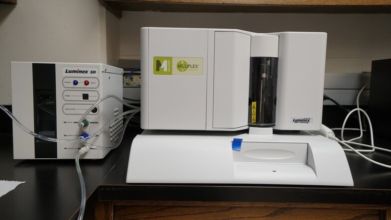
MILLIPLEX LUMINEX L200 ANALYZER

Milliplex Analyzer developed by Luminex uses multiplex xMAP bead technology that allows for teh simultaneous analysis of a broad selection of analytes (e.g. cytokines, hormones and adipokines) across a wide range of disease states, including metabolic disease, inflammation, neurodegenerative disease, toxicity, cancer and more.

Key Features:

Flow-Based Detection: Powered by xMAP® Technology, which uses a flow cytometry approach to identify and quantify individual microsphere sets, ensuring accurate multiplex results.

Versatile Multiplexing: Measures up to 100 targets in a single reaction, allowing users to conserve time, samples, and reagents. Compatibility with both magnetic (MagPlex®) and polystyrene microspheres offers flexibility for diverse assay needs.

High Throughput and Speed: Reads a 96-well plate in about 45 minutes (up to 12,800 tests per hour).

Broad Dynamic Range and Sensitivity: Greater than 3.5 logs dynamic range to detect low- to high-abundance targets. Sensitivity of fewer than 1,000 fluorochromes per microsphere supports the detection of subtle expression differences. 

Image

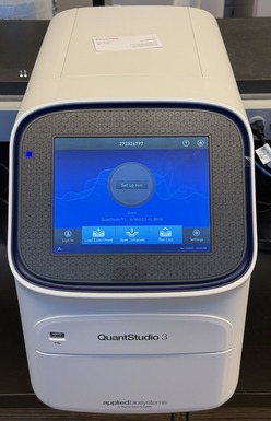
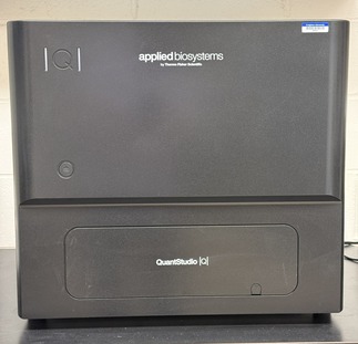
QUANTSTUDIO 3 REAL-TIME PCR

The Applied Biosystems™ QuantStudio™ 3 Real-Time PCR System is an accessible, high-performance benchtop instrument designed to empower researchers of all experience levels. Combining intuitive software, interactive touch-screen functionality, and secure connectivity via Thermo Fisher Connect, it enables quick setup, rapid data analysis, and easy collaboration.

Key Features:

Easy-to-use software and touch screen: An interactive interface and large, icon-driven touch screen guide you step-by-step through experiment setup and data analysis. Preoptimized protocol templates help eliminate guesswork, reducing time spent on training and troubleshooting.

Precise and reliable performance: With the ability to detect as little as 1.5-fold differences in target quantity in singleplex reactions and a broad dynamic range (up to 10 logs), the QuantStudio 3 system delivers the accuracy you need for gene expression, SNP genotyping, and other genomic applications.

Flexible experiment design: The system supports both probe-based (e.g., TaqMan®) and intercalating dye (e.g., SYBR™ Green) chemistries. Four optical channels allow for multiplexing of up to four targets in a single reaction, saving time and resources.

Image
Picture 4

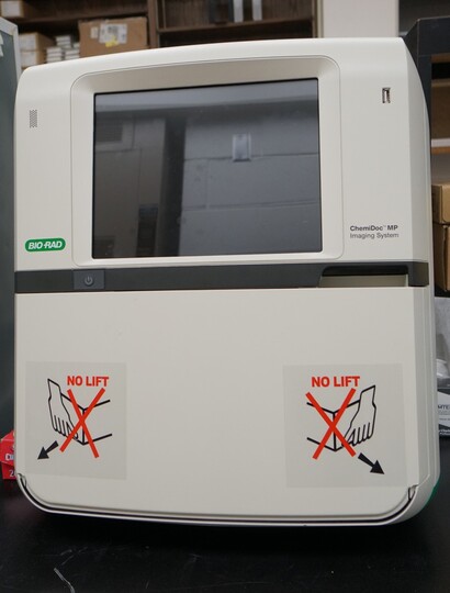
The ChemiDoc MP Imaging System is a full-feature instrument for imaging and analyzing gels and western blots.  It is designed to address multiplex fluorescent western blotting, chemiluminescence detection, general gel documentation applications, and stain-free technology imaging needs.  Features include:

  • All-in-one flexible imaging - get precise, reproducible fluorescent, chemiluminescence, and colorimetric gel and blot detection, analysis and documentation in a single system
  • Multiplex fluorescent western blotting - detect up to three proteins simultaneously and eliminate the need for stripping and reprobing
  • Ease of use - features include automatic selection of optimal light source by application, auto focus, auto exposure, and preview features ensure optimal images.  Learn in minutes with on screen help.
  • Stain-free protein normalization - stain-free imaging permits the normalization of bands to total protein in both in gels and blots; eliminates the need for housekeeping proteins
  • Sample Trays and Smart Tray Technology - three sample trays available to cover diverse imaging applications. Smart Tray Technology automatically recognizes the application-specific trays and adjusts imaging parameters and software options accordingly
Image


QUANTSTUDIO ABSOLUTEQ DIGITAL PCR

Elevate your research with the QuantStudio™ Absolute Q™ Digital PCR System—a powerfully simple platform that integrates all steps of digital PCR into a single instrument. Proprietary microfluidic array plate (MAP) technology ensures over 95% of your sample is accurately measured, delivering precise, sensitive, and absolute quantification without the need for standard curves. From genomics, oncology to infectious disease, this versatile system is designed to accelerate your discoveries with minimal hands-on time and streamlined data analysis.

Key Features:

Single-Instrument Workflow: Combine reaction digitization, thermal cycling, and data acquisition in one compact system.

Microfluidic Array Plate Technology: Achieve consistent sample partitioning, with less than 5% dead volume and up to 20,000 micro-chambers per reaction.

High Sample Utilization: Analyze over 95% of your sample to boost sensitivity and accuracy, ensuring maximum data recovery.

Four-Color Multiplexing: Detect up to four targets in a single run, saving time and reagents while expanding your application range.

Broad Application Range: Ideal for oncology, reproductive health, infectious disease, inherited disease, gene expression, copy number variation, and more.

User-Friendly Software: Easily set up plate layouts and thermal conditions, then visualize results in clear, exportable formats for downstream analysis.

Image

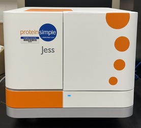
JESS CAPILLARY WESTERN

Meet Jess—the fully automated, next-generation protein analysis platform that brings together Western blotting into a single, easy-to-use system. Designed to eliminate tedious steps and human error, Jess streamlines protein separation, immunodetection, and data analysis into an efficient, three-hour workflow. Simply load your samples and reagents,  and let her handle the rest. Her high-sensitivity chemiluminescent and NIR/IR fluorescence modules capture picogram-level signals, giving you reliable, publication-ready results without the guesswork.

Whether you’re monitoring protein expression, running quantitative immunoassays, or comparing multiple targets, Jess maximizes your data output with minimal sample input—perfect for precious or low-abundance samples. 

Key Features:

Full Automation: Jess handles protein separation, immunodetection, and data analysis—all in a single run—minimizing hands-on time and reducing user-dependent variability.

High Sensitivity: Achieve picogram-level detection with chemiluminescence or capitalize on superior fluorescence performance with NIR/IR modules for low-abundance targets.

Efficient Workflows: Run up to 25 samples per cartridge, and analyze results in as little as three hours.

Multiplex and Normalization: Detect multiple proteins simultaneously and use built-in total protein or fluorescent normalization to ensure consistent, reproducible results.

Streamlined Data Analysis: Easy-to-use software delivers both qualitative (Western blot–like) and quantitative (ELISA-like) insights, simplifying protein sizing, concentration measurements, and comparative analyses.

RePlex Capability: Quickly swap between different immunoprobes or total protein detection in a single run, maximizing data acquisition from limited or precious samples.

Image

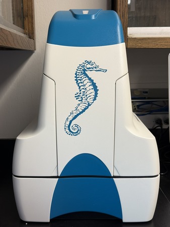
AGILENT SEAHORSE XFe24 FLUX ANALYZER

Experience a deeper understanding of cellular metabolism with the Agilent Seahorse XFe24 Analyzer—the trusted platform for real-time measurement of mitochondrial respiration and glycolysis in live cells. This high-sensitivity, 24-well system enables precise metabolic assessment of diverse samples, including islets, organoids and small organisms such as zebrafish larvae, C. elegans that require a larger well format. By simultaneously monitoring oxygen consumption and extracellular acidification, the Seahorse XFe24 delivers critical insights into how cells generate and expend energy. 

The Seahorse XFe24 Analyzer simplifies everything from routine assays to advanced metabolic investigations, helping researchers uncover the mechanisms behind disease, immunity, cancer, and more. Whether you’re optimizing new treatments or exploring fundamental cellular processes, the Seahorse XFe24 puts the power of real-time metabolic data at your fingertips—empowering you to make breakthrough discoveries faster and with greater confidence.

Key Features:

24-Well Format: Perfect for working with specialized cell types, including islets and small organisms, that require a larger well size than traditional microplate formats.

Real-Time Metabolic Analysis: Monitors both oxygen consumption and extracellular acidification simultaneously, providing a complete picture of mitochondrial respiration and glycolysis in live cells.

Automated Compound Injections: Allows up to four sequential reagent additions per well, enabling multi-step protocols and reducing manual handling for greater reproducibility.

High Sensitivity: Detects metabolic activity in as few as 5,000 cells per well, making it suitable for rare or precious cell samples.

Flexible Experimental Design: Compatible with both adherent and suspension cells, as well as isolated mitochondria, for diverse applications across research areas like cancer, immunology, and metabolic disorders.

Robust Data Analysis Software: Delivers intuitive, automated calculations of key parameters—such as basal respiration, ATP-linked respiration, and spare respiratory capacity—streamlining the path from raw data to meaningful insights.


*Directed by Sarath Vijayakumar

We offer multiple advanced research microscopes that are available for imaging-based experiments.  We also provide a selection of sophisticated software packages that can be utilized for image analysis.  Please inquire if you have specific questions about any of these instruments or about gaining access to them.  We are happy to provide training as well as to consult with users about their experimental and analysis needs.  New users must receive official training and sign a user agreement prior to gaining access to the facility's tools or its reservation systems.

Instrumentation:

The Advanced Imaging Core hosts three confocal laser scanning microscopes from Zeiss.  All three microscopes offer DIC imaging capabilities and popular objective magnifications (10X, 20X, 40X, 63X). The inverted LSM 700 offers four excitation wavelengths (405, 488, 555, 639 nm), while our upright LSM 710 has 7 laser lines (405, 458, 488, 514, 561, 594, 633), 2 PMT’s, and a 32-channel detector.  The facility also has a Zeiss LSM 980 with Airyscan, which offers high resolution, improved sensitivity, and capabilities for live cell imaging.  Finally, the AVT- Advanced Imaging Core manages a Zeiss PALM CombiSystem, which enables users to perform microdissection and micromanipulation of samples on standard glass slides within a single microscope.  These microscopes can be found in rooms 407, 332, and 312 of the Criss Complex.


Image
Zeiss

The Advanced Imaging team also manages several specialized microscopes associated with CU-IBIF, Creighton University’s Integrated Biomedical Imaging Facility, located in rooms 324, 325, 326C, 376, and 382A of the Criss Complex.

First is an inverted Nikon Ti-2 confocal microscope with a Yokogawa spinning disk, a Hamamatsu Orca Flash camera, and an incubated stage.  This microscope is capable of fast full-frame imaging, and it can perform Z-stacks at rates 10 times faster than a typical point scanning confocal.  This microscope is equipped with Live Super-Resolution, which enables the high-speed imaging that is required for live cell experiments.  It is also an ideal choice for extended time-lapse imaging experiments.

Next is an upright Leica SP8 confocal microscope that is suitable for live animal imaging and can support accessories for electrophysiology measurements.  In addition to visible-range lasers, it has a tunable, pulsed, near-infrared Ti:S laser for multiphoton excitation of UV/Visible fluorophores and second harmonic imaging. Non-descanned Super HyD detectors and a Becker and Hickl SPC 830 Time Correlated single photon counting system enable fluorescence lifetime imaging, fluorescence correlations spectroscopy, and lifetime-based FRET techniques. The Leica has spectral detectors that enable spectral imaging.

Image
Nikon and Leica

Third is a home-built Total-Internal Reflection Fluorescence Microscope.  This instrument offers a through-the-prism beam path to reduce noise from scattered laser light, and it incorporates three objective options (10X, 20X, 40X) and four laser lines (405, 488, 552, and 647 nm).  Images are captured with an Andor iXon EMCCD scientific camera.

The team also manages an inverted ImageXpress Micro 4 widefield microscope from Molecular Devices, equipped with an incubation chamber.  The ImageXpress enables users to conduct preprogrammed experiments over extended time periods at high-throughput.  In addition, CU-IBIF is also the home to two other widefield microscopes for basic imaging needs.

Image
TIRF and IMAGEXPRESS

Software Packages Available:

The Advanced Imaging Core provides a selection of analysis tools for the THC and users of our facility.  We have two workstations that run the Imaris Microscopy Image Analysis software, which is helpful in processing 3D and 4D images.  A standalone workstation is available for utilizing Nikon’s NIS Elements and Leica’s LAS X software.  Users interested in knowing more about these software packages and accessing them are asked to reach out to the team.  We are happy to discuss your image processing needs with you, and we have experience in using a variety of other programs as well for analysis and processing, including ImageJ, MATLAB, Python, and R.


The Mass Spectrometry Core offers two mass spectrometers and provides a selection of software to be utilized for the analysis of mass spectrometry-based experiments. Current capabilities include mass determination, proteomics, and absolute quantitation of small molecules. Please inquire with Molly McDevitt (mollymcdevitt@creighton.edu) if you have specific questions regarding the capabilities of the Mass Spectrometry Core. New users must sign a user agreement prior to running samples. At this time, no training for the individual use of instruments will be provided. However, we will happily provide software training so users can complete their own analysis of any mass spectrometry data, if desired.

Instrumentation

The Mass Spectrometry Core houses two mass spectrometers. Both can be found in Criss II Room 303.

Image
Bruker

The Bruker ultrafleXtreme is a MALDI-TOF/TOF mass spectrometer with resolving power up to 40,000 over a wide range of masses and up to 1 ppm mass accuracy. It is equipped with both MS and MS/MS capabilities, exhibiting high data acquisition speeds for both. It is ideal for mass determination and structure identification for a variety of biomolecules, including proteins, peptides, lipids, and glycans.

Image
Thermo

The Thermo Q Exactive is a hybrid quadrupole Orbitrap mass spectrometer which combines high-performance quadrupole precursor selection with high-resolution, accurate mass (HR/AM) Orbitrap detection. It boasts a resolving power of up to 140,000 FWHM and mass accuracy below 1 ppm. Fast scan speeds, the HR/AM capabilities, the capacity for MS/MS, and versatility of the instrument make it ideal for the identification and quantitation of small molecules, peptides, and proteins.

In addition to the two mass spectrometers, Criss II Room 303 is also home to two different liquid chromatography (LC) systems. Used in conjunction with the Thermo Q Exactive, both LC systems enable separation of analytes prior to analysis, reducing sample complexity and resulting in lower detection limits.

Image
Thermo

The Thermo Vanquish Flex Binary UHPLC system is comprised of a biocompatible, binary, high-pressure gradient mixing pump, an autosampler, and a heated column compartment. With a pressure limit of 1000 bar and flow rate range of 50 µL – 8 mL/min, it is suited for a variety of analyses, including absolute quantitation of small molecules, lipidomics, and metabolomics.

Image
Waters ACQUITY M-Class

The Waters ACQUITY M-Class Chromatography System provides nanoflow technology with the straightforward setup found in an analytical-scale UPLC system.  Nanoflow technology, adds increased sensitivity compared to analytical-scale UPLC systems and is ideal for analyses in which sample is limited. With a pressure limit of 15,000 psi and capable of flow rates as low as 200 nL/min, it is best suited for bottom-up proteomics, including data dependent acquisition, targeted analysis, and affinity-purification mass spectrometry.  

Software Packages:

The Mass Spectrometry Core provides a selection of analysis tools for users of our facility:

· fleXAnalysis

· Xcalibur Software Package

· TraceFinder 4.1

· Proteome Discoverer 3.1

For users that are interested in doing their own data analysis, please reach out to the team and we will be happy to provide training.


*Directed by David Smith and assisted by Molly McDevitt

Image
Zebrafish

The Zebrafish services of the Auditory & Vestibular Technology core provide support for behavioral, developmental, and neurobiological studies. Researchers have access to fish housing, breeding, and use of several instruments specific for using zebrafish as a model organism. We can support researchers and facility users through any of the following services. Additional project-specific support for users can be arranged as needed.

  •  
    • Behavioral Analysis
    • Drug Screening
    • Genetic Manipulation
    • Zebrafish Imaging

When publishing a research paper that relied on equipment and used within the AVT Core Facility, please remember to acknowledge the core. Proper acknowledgment is critical for tracking research papers and meets the requirements of our funding agencies.  We recommend that you use the statement below which cites the grant and research ID number.  It should not count against a publication's word count.  You may simply copy and paste this content into the Acknowledgements section of your paper:

This research was partially conducted within the Auditory and Vestibular Technologyies Core Facility (AVT) at Creighton University, Omaha, NE (RRID:SCR_023866). This facility is supported by the Translational Hearing Center at Creighton University, Boys Town National Research Hospital and University of Nebraska Medical Center with CoBRE Award GM139762 from the National Institute of General Medical Science, a component of the National Institutes of Health. This investigation is solely the responsibility of the authors and does not necessarily represent the official views of any supporting institution.